


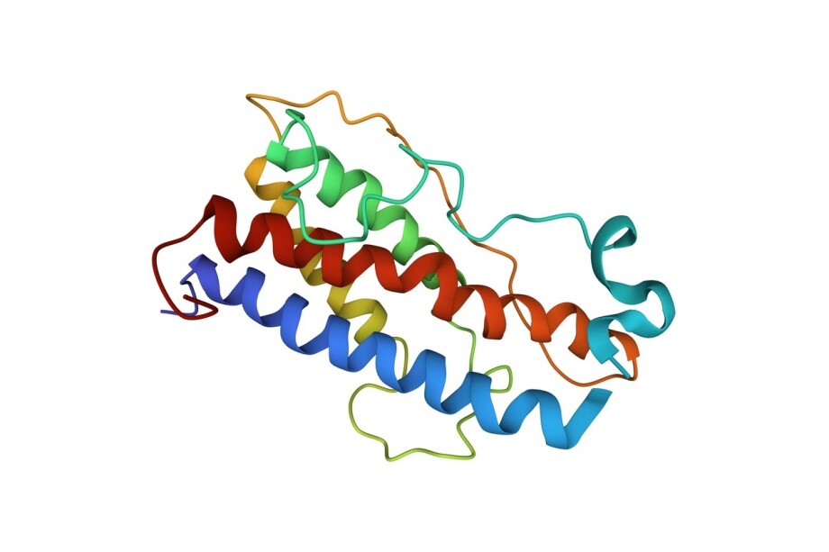
Tesamorelin and Sermorelin are posited to be synthetic research peptides designed to interact with growth hormone-releasing hormone (GHRH) receptors on anterior pituitary gland cells. Both molecules are classified as GHRH analogs and are studied in laboratory settings for their potential to stimulate endogenous growth hormone (GH) synthesis, pulsatile secretion, and downstream insulin-like growth factor-1 (IGF-1) signaling.
Although they share the same primary receptor target on pituitary somatotroph cells, Tesamorelin and Sermorelin differ in amino acid chain length, terminal chemical modifications, resistance to enzymatic cleavage, and the apparent magnitude of their downstream anabolic and catabolic signaling. Researchers also frequently compare both GHRH analogs against growth hormone secretagogues (GHSs) such as Ipamorelin, which engages a separate pituitary receptor pathway.
What Are Tesamorelin and Sermorelin?
Sermorelin and Tesamorelin are considered to be structural derivatives of endogenous GHRH, but may represent two distinct approaches to peptide design and stability. Sermorelin, also known as GRF(1–29)-NH₂ or GHRH(1–29)-NH₂, appears to be a truncated synthetic peptide composed of 29 amino acids corresponding to the first 29 N-terminal residues of the native 44-amino-acid GHRH sequence. Research by Clark et al. indicates that Sermorelin carries a C-terminal amidation and retains the biological activity associated with the N-terminal core of full-length GHRH, suggesting that the first 29 amino acids may be sufficient for GHRH receptor recognition and activation.(1)
While the C-terminal amidation is posited to help preserve a receptor-compatible conformation and slow certain forms of carboxy-terminal degradation, Sermorelin leaves the N-terminal Tyr¹-Ala² bond unmodified, and the peptide remains susceptible to rapid cleavage by dipeptidyl peptidase-IV (DPP-IV), yielding a half-life of approximately 10–12 minutes.
On the other hand, Tesamorelin apparently retains the complete 44-amino-acid backbone of GHRH along with specific terminal modifications intended to slow enzymatic breakdown. According to Ferdinandi et al., Tesamorelin may also incorporate a trans-3-hexenoyl group attached to the terminal nitrogen of Tyr¹ at the N-terminus, while terminating in an amidated -Leu⁴⁴-CONH₂ sequence at the C-terminus.(2) This N-terminal trans-3-hexenoyl moiety appears to sterically hinder DPP-IV from cleaving the Tyr¹-Ala² dipeptide, which would otherwise rapidly deactivate the molecule. Research by González-Sales et al. suggests that these structural protections may extend Tesamorelin’s half-life to roughly 25–40 minutes, compared with roughly 7–10 minutes for unmodified GHRH, and may support elevated GH synthesis from pituitary cells within 30–60 minutes of exposure.(3)
Differences in Mechanism of Action Between Tesamorelin vs Sermorelin
Both Tesamorelin and Sermorelin appear to act primarily by binding to the growth hormone-releasing hormone receptor, a class B G-protein-coupled receptor expressed on the surface of anterior pituitary somatotroph cells. Research by Culhane et al. suggests that peptide ligands may activate class B G-protein-coupled receptors through a two-domain mechanism in which the C-terminal region of the peptide initially associates with the receptor’s extracellular domain, positioning the N-terminal region to engage the juxtamembrane and transmembrane domains.(4) The authors suggest that “such binding leads to conformational changes in the receptor’s cytoplasmic domain to [activate] a G protein to trigger a downstream signaling process.”
Research by Halmos et al. indicates that activated GHRH receptors may preferentially couple to the Gαs subunit when presented with an intact three-dimensional ligand conformation.(5,6) Once either peptide successfully couples GHRH receptors to Gαs, activated Gαs appears to stimulate membrane-bound adenylyl cyclase, increasing intracellular cyclic adenosine monophosphate (cAMP) and activating protein kinase A (PKA).
Research by Takei et al. suggests that cAMP and intracellular calcium ions (Ca²⁺) may cooperate to regulate hormone exocytosis from pituitary cells, as GHRH receptor-driven depolarizing currents promote the opening of voltage-dependent Ca²⁺ channels.(7) The resulting influx of Ca²⁺ appears to trigger the fusion of GH-containing secretory vesicles with the plasma membrane. In parallel, Halmos et al. note that cAMP-PKA signaling may phosphorylate nuclear transcription factors such as cAMP response element-binding protein (CREB), potentially supporting GH gene transcription and the replenishment of intracellular hormone stores alongside acute granule release.(6)
Do Tesamorelin and Sermorelin Stimulate GH Pulses and IGF-1?
Laboratory evaluations suggest that both Tesamorelin and Sermorelin may stimulate pulsatile GH secretion from pituitary cells and upregulate downstream IGF-1 production. Yet, Tesamorelin may be associated with larger sustained shifts across 12-hour and multi-week observation windows. In studies of Sermorelin, the observed GH response appears tightly linked to the duration of the measurement interval. Vittone et al. reported that exposure to Sermorelin may increase mean 12-hour GH concentrations from approximately 1.1 ± 0.9 µg/L to around 2.2 ± 1.9 µg/L, while integrated 12-hour GH output rose approximately twofold from 1,114 ± 931 to 2,032 ± 1,728 µg·min/L.(8) When Khorram et al. evaluated a closely related [Nle²⁷]GHRH(1–29)-NH₂ sequence across shorter windows, the most pronounced secretory burst appeared within the first two hours of observation, during which integrated GH levels rose roughly fivefold to sixfold from 200–300 µg·L⁻¹·min to 1,100–1,600 µg·L⁻¹·min.(9)
Across repeated exposure, Khorram et al. reported that this pulsatile GH output was accompanied by an apparent increase in downstream IGF-1 levels of approximately 27–28%. Tesamorelin appears to possess a broader potential for stimulating both pulsatile and basal GH output, as well as a more pronounced rise in IGF-1. Research by Stanley et al. suggests that Tesamorelin may promote an overall increase in GH secretion driven by both elevated basal GH output and a larger average pulse area.(10) Specifically, their analysis indicated that Tesamorelin increased overnight 12-hour GH area under the curve (AUC) by 366 µg/L·h, representing an overall rise of roughly 69%, while average GH pulse area rose by approximately 55% without altering endogenous pulse frequency.
In the same investigation, Tesamorelin exposure was linked to an apparent upregulation of IGF-1 levels by roughly 122% from baseline, while preserving peripheral insulin sensitivity as assessed by euglycemic clamp M-values. Consistent with its stronger IGF-1 response, laboratory evaluations by Adrian et al. suggest that Tesamorelin exposure may be associated with significant increases in skeletal muscle density and lean muscle cross-sectional area reaching up to +12.1% above baseline.(11)
How Tesamorelin and Sermorelin May Interact with Visceral Fat Cells?
Neither Tesamorelin nor Sermorelin is thought to act directly on adipocytes, but rather indirectly via elevated pituitary GH output and subsequent GH receptor signaling on adipose tissue. Researchers such as Kopchick et al. have suggested that “GH [may interact with] adipose tissue in a depot-specific manner and [interacts with] other features of adipose tissue”.(12) This depot selectivity is posited to reflect a two- to threefold higher density of GH receptors on visceral fat cells compared with subcutaneous fat cells, allowing visceral adipocytes to mount an amplified catabolic response to rising pulsatile GH levels.
When GH binds to adipocyte GH receptors, downstream signaling may promote PKA-mediated phosphorylation of hormone-sensitive lipase (HSL) at Ser563/660 and recruitment of CGI-58 to activate adipose triglyceride lipase, accelerating the hydrolysis of stored triglycerides into free fatty acids and glycerol for β-oxidation. Simultaneously, activation of the JAK/STAT pathway may upregulate genes linked to lipid oxidation and uncoupling protein 1 (UCP1) while suppressing lipogenic enzymes that promote new lipid storage.
Experimental data on Tesamorelin indicates that its downstream signaling may reduce average visceral fat cell volume by approximately 15% without depleting subcutaneous fat depots.(10,12) Furthermore, research by Machado et al. suggests that heightened GH signaling may upregulate low-density lipoprotein (LDL) receptor expression on hepatic cells, while lower visceral lipid flux may reduce hepatic particle output, potentially corresponding to reductions of 15–20% in triglycerides and 5–10% in LDL cholesterol alongside a ~5% increase in HDL levels.(13)
How Does Ipamorelin Differ from Both Tesamorelin and Sermorelin?
While Tesamorelin and Sermorelin are both structural GHRH analogs that act through pituitary GHRH receptors, Ipamorelin belongs to a separate pharmacological class known as growth hormone secretagogues (GHSs) and operates through a distinct receptor and intracellular signaling cascade.
Structurally, Ipamorelin is a synthetic pentapeptide with the sequence Aib-His-D-2-Nal-D-Phe-Lys-NH₂, making it much shorter than either the 29-amino-acid Sermorelin or the 44-amino-acid Tesamorelin. Rather than mimicking endogenous GHRH, Ipamorelin functions as a selective mimetic of ghrelin and binds to the growth hormone secretagogue receptor type 1a (GHS-R1a) on pituitary somatotroph cells. Foundational research by Raun et al. suggested Ipamorelin may be the first selective GHS-R1a agonist capable of stimulating GH release with a selectivity profile comparable to GHRH itself, suggesting no statistically significant elevation in adrenocorticotropic hormone (ACTH), cortisol, prolactin, or luteinizing hormone even at concentrations far above those required for GH stimulation.(14)
At the intracellular level, Ipamorelin’s engagement of GHS-R1a triggers a signaling route distinct from the Gαs/cAMP/PKA pathway shared by Tesamorelin and Sermorelin. According to Yin et al., activated GHS-R1a receptors predominantly couple to Gq/11 proteins to stimulate phospholipase C (PLC).(15) PLC cleaves membrane phosphatidylinositol 4,5-bisphosphate (PIP₂) into inositol 1,4,5-trisphosphate (IP₃) and diacylglycerol (DAG). IP₃ subsequently promotes the rapid release of Ca²⁺ from intracellular endoplasmic reticulum stores, while DAG activates protein kinase C (PKC), driving depolarizing calcium influx and the exocytosis of GH-containing vesicles. In addition, GHS-R1a activation in hypothalamic cell networks may partially attenuate somatostatin tone, lessening the primary inhibitory brake on somatotroph GH secretion.
Pharmacokinetic and pharmacodynamic modeling by Gobburu et al. suggests that Ipamorelin exhibits a terminal half-life of roughly two hours, which is longer than the reported half-life of either Sermorelin at 10–12 minutes or Tesamorelin at 25–40 minutes.(16) In that evaluation, Ipamorelin induced a single, acute episode of GH release that peaked at approximately 0.67 hours, or about 40 minutes, before declining exponentially toward baseline. Because Ipamorelin acts via the Gq/PLC/IP₃ pathway while Tesamorelin and Sermorelin act via the Gαs/cAMP/PKA pathway, researchers view Ipamorelin as mechanistically distinct from, and potentially complementary to, both GHRH analogs.
Frequently Asked Questions
What is the difference between Tesamorelin and Sermorelin?
Both peptides are synthetic GHRH analogs, but they differ in chain length, chemical protection, and signaling potency. Sermorelin is a truncated 29-amino-acid fragment of native GHRH with a short half-life of about 10–12 minutes. Tesamorelin retains the full 44-amino-acid GHRH sequence plus an N-terminal trans-3-hexenoyl modification, extending its half-life to roughly 25–40 minutes and supporting more pronounced increases in GH pulsatile output.
How does Ipamorelin compare with Tesamorelin?
Tesamorelin appears to drive a broader, more sustained increase in endogenous GH synthesis via GHRH receptors, raising 12-hour GH output by roughly 69% and average GH pulse area by about 55% alongside a ~122% rise in IGF-1. Ipamorelin acts on ghrelin receptors (GHS-R1a) to trigger a single, acute burst of GH synthesis that peaks after about 40 minutes, reaching a modeled maximal GH production rate of 694 mIU/L/h before rapidly declining toward baseline.
How does Sermorelin compare with Ipamorelin?
Sermorelin stimulates GHRH receptors to produce an early fivefold to sixfold surge in GH synthesis over the first two hours and an approximately twofold increase in integrated GH output over 12 hours. Ipamorelin stimulates GHS-R1a receptors to induce an acute, single-episode GH spike peaking near 40 minutes without significantly altering ACTH or cortisol, and because the two peptides act on separate pituitary receptors, their combined exposure is posited to amplify GH synthesis beyond the sum of either peptide alone.
Can you combine Sermorelin and Tesamorelin?
No published laboratory studies appear to have evaluated Sermorelin and Tesamorelin in combination. From a receptor-signaling perspective, combining Sermorelin and Tesamorelin is generally considered mechanistically redundant because both molecules are GHRH analogs that compete for the same binding pocket on pituitary GHRH receptors.
NOTE: These products are intended for laboratory research use only. This peptide is not intended for personal use. Please review and adhere to our Terms and Conditions before ordering.
References:
- Clark RG, Robinson ICAF. Growth induced by pulsatile infusion of an amidated fragment of human growth hormone releasing factor in normal and GHRF-deficient rats. Nature. 1985;314(6008):281–283. https://doi.org/10.1038/314281a0
- Ferdinandi ES, Brazeau P, High K, Procter B, Fennell S, Dubreuil P. Non-clinical pharmacology and safety evaluation of TH9507, a human growth hormone-releasing factor analog. Basic Clin Pharmacol Toxicol. 2007;100(1):49–58. https://doi.org/10.1111/j.1742-7843.2007.00008.x
- González-Sales M, Barrière O, Tremblay PO, et al. Population pharmacokinetic and pharmacodynamic analysis of tesamorelin in HIV-infected patients and healthy subjects. J Pharmacokinet Pharmacodyn. 2015;42(3):287–299. https://doi.org/10.1007/s10928-015-9416-2
- Culhane KJ, Liu Y, Cai Y, Yan ECY. Transmembrane signal transduction by peptide hormones via family B G protein-coupled receptors. Front Pharmacol. 2015;6:264. https://doi.org/10.3389/fphar.2015.00264
- Zhou F, Zhang H, Cong Z, et al. Structural basis for activation of the growth hormone-releasing hormone receptor. Nat Commun. 2020;11(1):5205. https://doi.org/10.1038/s41467-020-18945-0
- Halmos G, Szabo Z, Dobos N, Juhasz E, Schally AV. Growth hormone-releasing hormone receptor (GHRH-R) and its signaling. Rev Endocr Metab Disord. 2025;26(3):343–352. https://doi.org/10.1007/s11154-025-09952-x
- Takei T, Yasufuku-Takano J, Takano K, Fujita T, Yamashita N. Effect of Ca2+ and cAMP on capacitance-measured hormone secretion in human GH-secreting adenoma cells. Am J Physiol. 1998;275(4):E649–E654. https://doi.org/10.1152/ajpendo.1998.275.4.E649
- Vittone J, Blackman MR, Busby-Whitehead J, et al. Effects of single nightly injections of growth hormone-releasing hormone (GHRH 1-29) in healthy elderly men. Metabolism. 1997;46(1):89–96. https://doi.org/10.1016/S0026-0495(97)90174-8
- Khorram O, Laughlin GA, Yen SSC. Endocrine and metabolic effects of long-term administration of [Nle27]growth hormone-releasing hormone-(1-29)-NH2 in age-advanced men and women. J Clin Endocrinol Metab. 1997;82(5):1472–1479. https://doi.org/10.1210/jcem.82.5.3943
- Stanley TL, Chen CY, Branch KL, Makimura H, Grinspoon SK. Effects of a growth hormone-releasing hormone analog on endogenous GH pulsatility and insulin sensitivity in healthy men. J Clin Endocrinol Metab. 2011;96(1):150–158. https://doi.org/10.1210/jc.2010-1587
- Adrian S, Scherzinger A, Sanyal A, et al. The growth hormone releasing hormone analog, tesamorelin, decreases muscle fat and increases muscle area in adults with HIV. J Frailty Aging. 2019;8(3):154–159. https://doi.org/10.14283/jfa.2018.45
- Kopchick JJ, Berryman DE, Puri V, Lee KY, Jorgensen JOL. The effects of growth hormone on adipose tissue: old observations, new mechanisms. Nat Rev Endocrinol. 2020;16(3):135–146. https://doi.org/10.1038/s41574-019-0280-9
- Machado MO, Hirata RD, Hirata MH, Hirszel P, Sellitti DF, Doi SQ. Growth hormone increases low-density lipoprotein receptor and HMG-CoA reductase mRNA expression in mesangial cells. Nephron Exp Nephrol. 2003;93(4):e134–e140. https://doi.org/10.1159/000070237
- Raun K, Hansen BS, Johansen NL, et al. Ipamorelin, the first selective growth hormone secretagogue. Eur J Endocrinol. 1998;139(5):552–561. https://doi.org/10.1530/eje.0.1390552
- Yin Y, Li Y, Zhang W. The growth hormone secretagogue receptor: its intracellular signaling and regulation. Int J Mol Sci. 2014;15(3):4837–4855. https://doi.org/10.3390/ijms15034837
- Gobburu JV, Agersø H, Jusko WJ, Ynddal L. Pharmacokinetic-pharmacodynamic modeling of ipamorelin, a growth hormone-releasing peptide, in human volunteers. Pharm Res. 1999;16(9):1412–1416. https://doi.org/10.1023/a:1018955126402
Available from Core Peptides
Supplied for laboratory research use only.
Related reading
- Sermorelin & Ipamorelin Blend: Research in Growth Hormone Modulation
- Sermorelin: Potential Interactions with Different Hormone-Producing Cells
- Sermorelin & GHRP-2 Synergistic Potential on Growth Hormone and IGF-1 Synthesis
- The Potential of Anabolism and Synergism in a Sermorelin & Ipamorelin Blend
- Sermorelin & GHRP-6: A Research Perspective on Dual-Pathway Pituitary Stimulation
- Sermorelin & GHRP-6 & GHRP-2 Blend: Anabolic Potential and Synergism
- Sermorelin’s Structure, Receptor Signaling, and Research Potential
- Tesamorelin Peptide: Potential Influence on the Hypothalamic–Pituitary–Somatotropic Axis
- Tesamorelin & Ipamorelin Blend: Synergistic Potential in Pituitary Cells
- Tesamorelin’s Potential for Anabolic and Catabolic Signaling in Cells