Insulin-like growth factor I (IGF-1) is considered to act as a crucial regulator in the mechanisms of physiological growth, primarily synthesized in response to stimulation by growth hormone. Among its notable variants, Mechano Growth Factor (MGF) emerges as a potential mediator involved in tissue growth and repair processes across diverse physiological systems, including muscle, tendon, cardiac, cerebral, and skeletal structures. The expression of MGF appears to dynamically respond to various physiological stimuli, orchestrating cellular proliferation and migration, which thereby, researchers speculate, may significantly influence tissue homeostasis and adaptive responses.(1)
The development of PEG-MGF (polyethylene glycol modified Mechano Growth Factor) peptides stemmed from the discovery of MGF.(2) PEG-MGF represents a synthetic peptide derived from the natural growth factor. Incorporating polyethylene glycol (PEG), a biocompatible polymer, into MGF peptides, is suggested by researchers to optimize their delivery and bioavailability. The synergistic action between MGF and has been researched due to the supposed inherent limitations associated with the rapid degradation of MGF.(3) The inclusion of polyethylene glycol (PEG) to peptides or proteins, commonly referred to as PEGylation, is also speculated to enhance water solubility and stability, as well as potentially decrease the molecule’s clearance through the kidneys, possibly resulting in an extended circulation time(3).
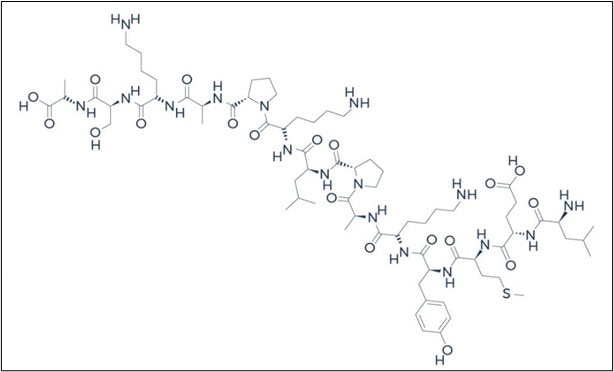
Fig. 1 – PEG-MGF Peptide Chemical Structure
Research
PEG-MGF Peptide and Skeletal Muscle Regeneration
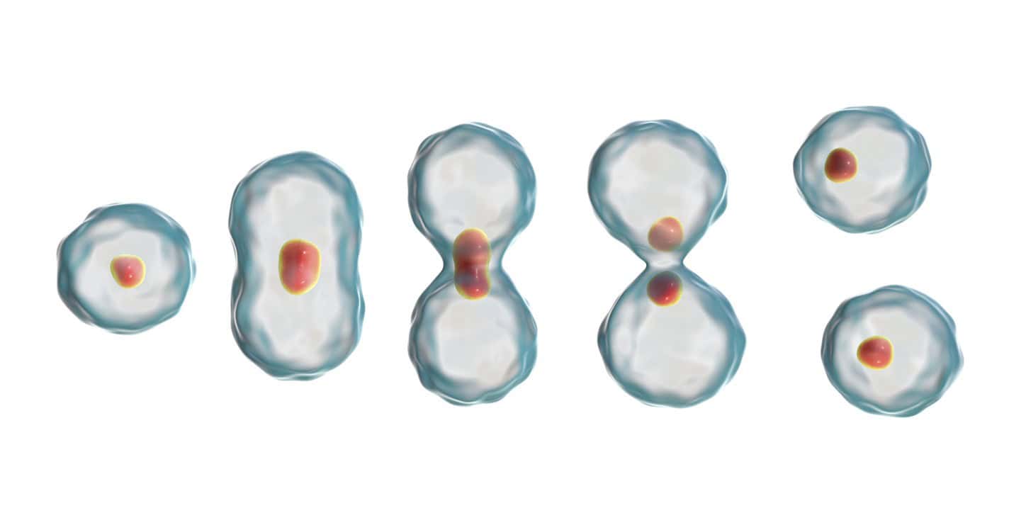
Skeletal muscle injuries are prevalent, and the process of regeneration following an injury is intricate. Studies(4) in murine models suggest exposure to mechano-growth factor (MGF) into injured muscles may confer protection by potentially mitigating inflammatory hormone expression and oxidative stress.(4)
Similarly, investigations(5) into the potential action of MGF on muscle inflammation following skeletal muscle injury suggest a possible regulatory role for MGF, which has spurred further research.
PEG-MGF Peptide and Cardiac Repair
Research conducted by the Department of Bioengineering at the University of Illinois indicates that PEG-MGF may exhibit inhibitory effects on programmed cell death in cardiac muscle subjected to hypoxia. Additionally, MGF appears to facilitate the recruitment of cardiac stem cells to the site of injury, thereby potentially aiding in cardiac regeneration and healing post myocardial infarction.(6)
Correspondingly, studies also suggest that localized exposure of PEG-MGF may contribute to enhanced cardiac function post myocardial infarction by attenuating pathological hypertrophy and promoting favorable hemodynamic outcomes.(7) Studies posit that the results indicate “that cardiac restricted administration of the MGF E-domain peptide using polymeric microstructures may [relevant to the prevention of] adverse remodeling of the heart and improve function following myocardial infarction.”
PEG-MGF Peptide and Bone Repair and Growth
Studies conducted in rabbits suggest that PEG-MGF exposure may expedite bone repair processes by potentially augmenting osteoblast proliferation, the cells responsible for bone mineralization. The reported healing outcomes in rabbits receiving PEG-MGF hint at the peptide’s capacity for bone repair mechanisms.(8)
PEG-MGF Peptide and Cartilage Protection
Research findings propose that MGF may potentially enhance chondrocyte function, considered crucial for maintaining cartilage integrity. Studies in murine models suggest that PEG-MGF peptide may facilitate the migration of chondrocytes from bone to cartilage.(9) Studies further suggest the “activity of the p38 MAPK pathway of NP cells cultured under mechanical overload was decreased by addition of MGF peptide”, suggesting that “MGF [may be] able to attenuate mechanical overload-induced NP cell apoptosis, and the p38 MAPK signaling pathway may be involved in this process.”
PEG-MGF Peptide and Dental Studies
In vitro studies conducted on periodontal ligament cells suggest that PEG-MGF may enhance the expression of ligament repair factors, such as MMP-1 and MMP-2, deemed essential for maintaining tooth stability within the bone. These findings suggest a potential for PEG-MGF peptide to aid in preserving natural teeth following damage.(10)
PEG-MGF Peptide and Neuroprotection
Multiple investigations(11) were undertaken utilizing murine models to elevate MGF levels, facilitating the examination of heightened MGF concentrations on brain cells. Among these studies, one involved the breeding of mice to constitutively overexpress MGF within the hippocampal region of the brain. The hippocampus is primarily associated with regulating neurogenesis. Studies report suggests that there was an overexpression of MGF, which is speculated to have led to elevated concentrations of BrdU, a recognized biological marker indicative of proliferative activity.
Research by Windebank and colleagues at the Mayo Clinic suggests that increasing MGF levels may potentially promote the growth of new brain cells in regions associated with brain renewal. Furthermore, higher MGF levels in transgenic mice appear to confer resistance to age-related brain damage and improved cognitive function, indicating a potential neuroprotective role for MGF in addressing age-related neuron loss and neurodegenerative disorders.
NOTE: These products are intended for laboratory research use only. This peptide is not intended for personal use. Please review and adhere to our Terms and Conditions before ordering.
References:
- Zabłocka, P. H.Goldspink, D.C. Górecki, “Mechano-Growth Factor : an important cog or a loose screw in the repair machinery?” Frontiers in Endocrinology, pp.1, 2012. https://www.ncbi.nlm.nih.gov/pmc/articles/PMC3485521/
- Hamley, I. W, “PEG−Peptide Conjugates.”American Chemical Society, pp.1543, 2014 https://centaur.reading.ac.uk/37092/
- Janssen, J. A., Hofland, L. J., Strasburger, C. J., van den Dungen, E. S., & Thevis, M. (2016). Potency of Full-Length MGF to Induce Maximal Activation of the IGF-I R Is Similar to Recombinant Human IGF-I at High Equimolar Concentrations. PloS one, 11(3), e0150453. https://www.ncbi.nlm.nih.gov/pmc/articles/PMC4798685/
- Liu X, Zeng Z, Zhao L, Chen P, Xiao W. Impaired Skeletal Muscle Regeneration Induced by Macrophage Depletion Could Be Partly Ameliorated by MGF Injection. Front Physiol. 2019 May 17;10:601. doi: 10.3389/fphys.2019.00601. PMID: 31164836; PMCID: PMC6534059. https://www.ncbi.nlm.nih.gov/pmc/articles/PMC6534059/
- Xu Q, Fang H, Zhao L, Zhang C, Zhang L, Tian B. Mechano growth factor attenuates mechanical overload-induced nucleus pulposus cell apoptosis through inhibiting the p38 MAPK pathway. Biosci Rep. 2019 Mar 28;39(3):BSR20182462. doi: 10.1042/BSR20182462. PMID: 30858307; PMCID: PMC6438874. https://www.ncbi.nlm.nih.gov/pmc/articles/PMC6438874/
- Doroudian G, Pinney J, Ayala P, Los T, Desai TA, Russell B. Sustained delivery of MGF peptide from microrods attracts stem cells and reduces apoptosis of myocytes. Biomed Microdevices. 2014 Oct;16(5):705-15. https://pubmed.ncbi.nlm.nih.gov/24908137/
- Peña JR, Pinney JR, Ayala P, Desai TA, Goldspink PH. Localized delivery of mechano-growth factor E-domain peptide via polymeric microstructures improves cardiac function following myocardial infarction. Biomaterials. 2015 Apr;46:26-34. doi: 10.1016/j.biomaterials.2014.12.050. Epub 2015 Jan 16. PMID: 25678113; PMCID: PMC4328136. https://www.ncbi.nlm.nih.gov/pmc/articles/PMC4328136/
- Deng M, Zhang B, Wang K, Liu F, Xiao H, Zhao J, Liu P, Li Y, Lin F, Wang Y. Mechano growth factor E peptide promotes osteoblasts proliferation and bone-defect healing in rabbits. Int Orthop. 2011 Jul;35(7):1099-106. doi: 10.1007/s00264-010-1141-2. Epub 2010 Nov 6. PMID: 21057789; PMCID: PMC3167400. https://www.ncbi.nlm.nih.gov/pmc/articles/PMC3167400/
- Xu Q, Fang H, Zhao L, Zhang C, Zhang L, Tian B. Mechano growth factor attenuates mechanical overload-induced nucleus pulposus cell apoptosis through inhibiting the p38 MAPK pathway. Biosci Rep. 2019 Mar 28;39(3):BSR20182462. doi: 10.1042/BSR20182462. PMID: 30858307; PMCID: PMC6438874. https://www.ncbi.nlm.nih.gov/pmc/articles/PMC6438874/
- Chen JT, Wang Y, Zhou ZF, Wei KW. [Mechano-growth factor regulated cyclic stretch-induced osteogenic differentiation and MMP-1, MMP-2 expression in human periodontal ligament cells by activating the MEK/ERK1/2 pathway]. Shanghai Kou Qiang Yi Xue. 2019 Feb;28(1):6-12. Chinese. PMID: 31080992. https://pubmed.ncbi.nlm.nih.gov/31080992/
- Alec Walker. Hearts and Minds of Mice and Men: Mechano Growth Factor a new tool in the battle against age-related neuron loss? 20 Jul 2017.