


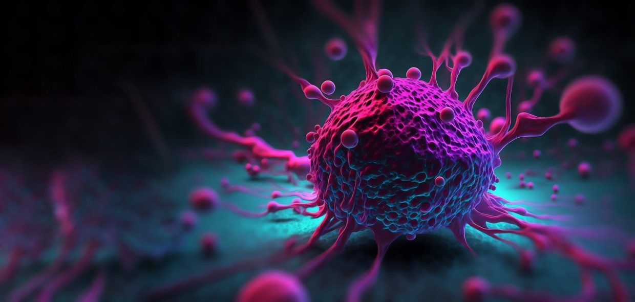
Мost laboratory work suggests that PNC-27 Peptide possibly binds HDM-2 present in the plasma membrane of transformed cells and then participates in transmembrane pore formation, leading to necrosis-like cell death that appears p53-independent in research models. Additional studies have posited secondary actions at mitochondrial membranes. Current research implications may include using PNC-27 peptide as a tool to probe HDM-2 localization in tumor cell membranes and studying tumor cell necrosis.
Research
PNC-27 Affinity to Proteins
Studies by researchers such as Sarafraz-Yazdi et al. suggest that PNC-27’s main mechanism may involve interaction with the oncoprotein HDM-2 via a p53‑derived binding sequence.(1) HDM-2’s normal role is considered to be the inactivation of p53’s suppressive role on tumor genes in the cell’s nucleus. In some tumor cells, HDM-2 may also be present on the membrane as a membrane-associated docking site.
The researchers also commented that there are “significant levels of HDM-2 in the membranes of a variety of cancer cells but not in the membranes of several untransformed cell lines”. This suggests that membrane localization of HDM-2 may, in some cases, be a hallmark of these transformed cells.
Binding studies using fluorescently labeled PNC-27 also suggest that this peptide associates with HDM-2 at the membrane in mammalian cancer cells. In contrast, in HDM-2–negative membranes, PNC-27 appears to diffuse inward without being retained at the surface. Moreover, these experiments support a causal link between membrane HDM-2 and necrosis induction.
The researchers commented that when full-length HDM-2 is forced to the membrane of otherwise resistant untransformed MCF-10-2A cells via a CAAX membrane-targeting motif, these cells become susceptible to PNC-27–induced LDH release and loss of viability. In contrast, cells expressing a membrane-localized HDM-2 variant lacking the p53/PNC-27–binding domain (residues 1–109) do not appear to have comparable sensitivity. This pattern suggests that the intact p53-binding pocket of membrane-resident HDM-2 may be required as a docking site.
Once bound, PNC-27 may retain its amphipathic helix–loop–helix conformation and, by remaining anchored within the bilayer, promote transmembrane pore formation. The consistent absence of caspase activation across these conditions is compatible with a necrosis-like, rather than apoptotic mode of death.
PNC-27 Activity in Mitochondria
Recent work by Krzesaj et al has proposed that PNC-27 might not be restricted to acting at the plasma membrane via HDM-2, but also “binds to the membranes of mitochondria, resulting in their disruption” once it has gained intracellular access.(2) In this study, the authors examined PNC-27–exposed MIA-PaCa-2 tumor cells using organelle-selective dyes and ultrastructural methods, building on the previously posited model of HDM-2–guided pore formation at the cell surface.
They suggest that the mitochondria of exposed cells apparently lose their capacity to retain mitotracker dye. In contrast, lysotracker staining remains largely preserved, which is interpreted as pointing to a relatively selective impairment of mitochondrial function in mammalian models.
Immuno-electron microscopy using gold-labeled anti-PNC-27 antibodies suggests that PNC-27 localizes not only to the plasma membrane but also to mitochondrial membranes under these conditions. This pattern is taken to imply that the peptide may transit, perhaps via its own pores or via damaged membranes, into the cytoplasm and then accumulate at mitochondria, where it possibly exerts additional membrane-active implications. In relation to its amphipathic helix-loop-helix structure, PNC-27 is posited to either form or expand pores in mitochondrial membranes.
The authors of these studies therefore outline a two-step model in which PNC-27 may first target membrane HDM-2 at the cell surface, inducing pore formation and early necrosis-like leakage, and then potentially extends its membrane-disruptive activity to mitochondria, further amplifying bioenergetic failure and cell death.
PNC-27 and Necrosis Induction in Different Cells
Research by Davitt et al. using a K562 cell model suggests the peptide may recognize membrane-associated HDM-2 on these types of tumor cells and position itself at their cellular surface.(3) Confocal microscopy in K562 cells suggested apparent co-localization of PNC-27 with HDM-2 as overlapping signals at the membrane, which the authors interpreted as data that these two components associate in situ.
Functionally, the study suggests that PNC-27 exposure may be followed by LDH release and near-complete loss of K562 cell viability, whereas caspase-3/7 activity apparently remained at baseline and classical apoptotic markers were not detected.
This may imply that PNC-27 possibly induces a necrosis-like mode of cell death linked to membrane permeabilization rather than apoptosis. The authors further suggest that this process is p53-independent, because K562 cells do not have p53 and yet still appear to undergo peptide-induced cell death.
By comparing tumor-derived cells to non-transformed controls, the study posits that susceptibility to PNC-27 correlates with the presence of HDM-2 in the plasma membrane. Cells lacking substantial membrane HDM-2, or expressing HDM-2 variants without the p53/PNC-27 binding domain, may not exhibit the same response, and forced expression of membrane-localized HDM-2 in otherwise resistant cells appears to render them vulnerable.
Research by Thadi et al. also suggests similar findings in U937, OCI-AML3, and HL-60 cells.(4) These cells also appear to express high levels of HDM-2 at the plasma membrane, whereas normal mononuclear cells may display only minimal surface HDM-2. This pattern is interpreted as indicating that aberrant membrane HDM-2 expression may function as a distinguishing feature between malignant and non-malignant hematopoietic cells.
After brief exposure, PNC-27 was once again suggested to reduce tumor cell viability in an apparently concentration-dependent fashion, markedly triggering LDH release into the supernatant, indicating necrosis. The study also suggests apoptosis markers such as Annexin V and caspase-3 activity remain at baseline in PNC-27–exposed cells, whereas classical apoptosis inducers produce robust Annexin V positivity and caspase-3 activation.
PNC-27 Selectivity Towards Tumor Cells
Research by Aguon et al. suggests that the aforementioned research conducted in laboratory settings involving selectivity assays is typically limited to a small panel of non-tumor cell lines, so they may underestimate heterogeneity in HDM‑2 membrane expression or ignore stressed and inflamed non-tumor cells.
Their research suggests that if PNC‑27 indeed promotes pore formation and necrosis by binding membrane HDM‑2, one concern is that endothelial cells, stromal cells, or regenerating epithelia might express enough HDM‑2 to also experience necrosis. Therefore, further research with a wide range of tumor cell lines and non-tumor cell lines should be conducted.
NOTE: These products are intended for laboratory research use only. This peptide is not intended for personal use. Please review and adhere to our Terms and Conditions before ordering.
References:
- Sarafraz-Yazdi E, Bowne WB, Adler V, Sookraj KA, Wu V, Shteyler V, Patel H, Oxbury W, Brandt-Rauf P, Zenilman ME, Michl J, Pincus MR. Anticancer peptide PNC-27 adopts an HDM-2-binding conformation and kills cancer cells by binding to HDM-2 in their membranes. Proc Natl Acad Sci U S A. 2010 Feb 2;107(5):1918-23. Epub 2010 Jan 11. PMID: 20080680; PMCID: PMC2836618.https://doi.org/10.1073/pnas.0909364107
- Krzesaj P, Adler V, Feinman RD, Miller A, Silberstein M, Yazdi E, Pincus MR. Anti-Cancer Peptide PNC-27 Kills Cancer Cells by Unique Interactions with Plasma Membrane-Bound hdm-2 and with Mitochondrial Membranes Causing Mitochondrial Disruption. Ann Clin Lab Sci. 2024 Mar;54(2):137-148. PMID: 38802154.
- Davitt K, Babcock BD, Fenelus M, Poon CK, Sarkar A, Trivigno V, Zolkind PA, Matthew SM, Grin’kina N, Orynbayeva Z, Shaikh MF, Adler V, Michl J, Sarafraz-Yazdi E, Pincus MR, Bowne WB. The anti-cancer peptide, PNC-27, induces tumor cell necrosis of a poorly differentiated non-solid tissue human leukemia cell line that depends on expression of HDM-2 in the plasma membrane of these cells. Ann Clin Lab Sci. 2014 Summer;44(3):241-8. PMID: 25117093.
- Thadi A, Lewis L, Goldstein E, Aggarwal A, Khalili M, Steele L, Polyak B, Seydafkan S, Bluth MH, Ward KA, Styler M, Campbell PM, Pincus MR, Bowne WB. Targeting Membrane HDM-2 by PNC-27 Induces Necrosis in Leukemia Cells But Not in Normal Hematopoietic Cells. Anticancer Res. 2020 Sep;40(9):4857-4867. doi: 10.21873/anticanres.14488. PMID: 32878773.
- Aguon PM, Aasen T, Distler ES, Mallin E. Experimental PNC-27 Therapy and Massive GI Hemorrhage: A Complication or Coincidence?: 1879. Official journal of the American College of Gastroenterology| ACG. 2017 Oct 1;112:S1035-6.