Biopeptide EL appears to share the sequence VGVAPG, which is also observed repeatedly in studies of key extracellular matrix proteins, such as elastin. Therefore, researchers have posited that the Biopeptide EL interacts with elastin-producing cells, such as fibroblasts. Interestingly, the peptide may mimic an excess of elastin, which may contribute to the stimulation of fibroblast migration and possibly proliferation. This may lead to a compensatory increase in other extracellular matrix proteins, such as collagen, hyaluronan, proteoglycans, fibronectin, laminin, and others. This hexapeptide structure of the Biopeptide EL is attached to a palmic acid fragment at the N-terminus. This palmitoylation is posited to support its penetration into the deeper layers of various dermal cell research models.
Research
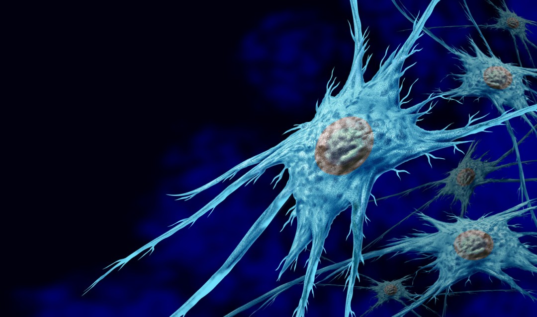
Biopeptide EL And Fibroblast Attraction
The research by Senior et al. suggests that the amino acid sequence VGVAPG in the Biopeptide EL may act as a chemotactic cue for fibroblasts and, to a lesser degree, monocytic cells.(1) In these research settings, fibroblasts were observed to migrate towards the Biopeptide EL vigorously with intensity that was roughly half to three-quarters of that evoked by a typical laboratory chemoattractant such as platelet-derived growth factor. Monocytes responded similarly but became refractory after prior exposure to elastin, implying a receptor-level desensitisation. Senior et al. also observed that the Biopeptide EL does not attract undifferentiated fibroblasts, but only mature and functional fibroblasts with the capacity to produce elastin.
The aforementioned chemotactic signal may also be neutralised by anti-elastin antibodies, according to reports by the researchers. Collectively, the data positions the Biopeptide EL as a potential tool for probing how dermal fibroblasts sense and move toward elastin-like cues. It may serve as a minimalist surrogate for the larger elastin molecule, providing a controllable, possibly receptor-specific signal to investigate migration, differentiation status, and extracellular matrix interactions in laboratory settings set up to observe dermal cell research models.
Biopeptide EL And Fibroblast Proliferation
Research by Kamoun et al. suggests Biopeptide EL may act as a mitogenic cue for cultured skin fibroblasts.(2) When experimental skin fibroblast cell cultures were exposed to the peptide, DNA synthesis and later cell counts rose to roughly double the values seen in peptide-free controls. A three- to five-day lag seemingly preceded the proliferative burst, and this delay shortened when the initial plating density was high. The authors posit that such density dependence may reflect autocrine or paracrine factors that accumulate as cultures near confluence.
In parallel, fibroblasts observed in the study gradually adopted a more elongated outline. They sometimes cluster around insoluble elastin fragments, implying that the Lipopeptide’s growth promoting actions might be accompanied—or even conditioned—by cytoskeletal rearrangements and cell–matrix tethering. Mechanistically, the peptide appears to interact with the 67 kDa elastin receptor that binds a repeated VGVAPG motif in tropoelastin.
Earlier work cited in the aforementioned article links receptor occupancy to G-protein–coupled activation of phospholipase C, inositol trisphosphate production, and calcium influx—pathways commonly associated with proliferation signals. For future in vitro dermatological research, the Biopeptide EL may help modulate fibroblast proliferation in three-dimensional dermal equivalents, wound-closure assays, or co-culture systems that explore epithelial–mesenchymal crosstalk.
Biopeptide EL And Elastin Synthesis
Research by Tajima et al. has confirmed previous research findings that the Biopeptide EL, and particularly its amino acid sequence “VGVAPG [may] stimulate skin fibroblast proliferation
and downregulate elastin expression”. The researchers also highlight that it may “downregulate elastin expression.” In parallel with its proliferative potential, the Biopeptide EL was suggested to consistently halve the levels of elastin mRNA while leaving collagen α1(I) transcripts unchanged, at least under the same exposure conditions.
The observed down-regulation was not believed to be related to mitigated cell count, as the fibroblast count was higher. Instead, the Biopeptide EL may have transcriptional or post-transcriptional control over the process of elastin synthesis. Researchers have proposed that this Biopeptide EL-driven repression in dermal fibroblasts may represent a negative feedback mechanism within elastin metabolism across species. Therefore, it may be posited that the observed increase in the number of fibroblasts may produce less elastin and more of other extracellular proteins when exposed to the Biopeptide EL.
Biopeptide EL And Collagen Breakdown
Research by Veiga et al. suggests that the Biopeptide EL may also act as a signal-type messenger, apparently moderating the inflammatory tone of cultured dermal cells.(4) In keratinocyte and fibroblast monolayers, the peptide has been suggested to curb interleukin-6 (IL-6) synthesis. This cytokine usually stays low but may surge within minutes after ultraviolet irradiation or other laboratory-specific stressors.(5) Lower IL-6 might, in turn, dampen the chemo-attraction of immune cells in co-cultures.
In part because recruited leukocytes generate reactive oxygen species that may trigger the transcription of matrix metalloproteinase (MMP) enzymes, the peptide is posited to mitigate the downstream burst of MMP activity, such as MMP-1 or MMP-9, and thereby slow the erosion of the collagen-rich matrix observed in cellular aging models. Indeed, some experiments in cellular aging models suggest that with 4 weeks of evaluation, the Biopeptide EL may support the firmness of dermal tissue in research models “by 20.0 % and increase skin tone by 33.0 % compared to a placebo.(4)
Biopeptide EL And Melanocytes
As noted by Veiga et al., the peptide may also support dermal pigment in research models, which might potentially be attributed to an additional specific receptor interaction. More specifically, a paper by Chang et al. suggests that melanocyte cell precursors may carry elastin-binding protein (EBP) receptors on their surface and that the Biopeptide EL may interact with them.(6) The cell cultures exposed to the Biopeptide EL also displayed more and longer dendrites, implying that the compound possibly promotes the cytoskeletal remodeling associated with pigment transfer.
Electron microscopy further revealed that melanosomes progressed from early stages to stage IV of maturation more readily after peptide administration. This pattern aligns with the biochemical observation that tyrosinase mRNA was upregulated in NCCmelan5 cells. In future research, the Biopeptide EL might be deployed to generate more pigmented, dendrite-rich melanocyte cultures. This may potentially provide researchers with a convenient system for probing pigment synthesis, organelle movement, and EBP-mediated signal cascades. It may also help model dermal melanocytosis in vitro, as it appears to recapitulate the juxtaposition of melanocyte dendrites with elastic structures.
You can find Lipopeptide for sale with 99% purity, on our website (available for research use only).
NOTE: These products are intended for laboratory research use only. This peptide is not intended for personal use. Please review and adhere to our Terms and Conditions before ordering.
References:
- Senior RM, Griffin GL, Mecham RP, Wrenn DS, Prasad KU, Urry DW. Val-Gly-Val-Ala-Pro-Gly, a repeating peptide in elastin, is chemotactic for fibroblasts and monocytes. J Cell Biol. 1984 Sep;99(3):870-4. doi: 10.1083/jcb.99.3.870. PMID: 6547961; PMCID: PMC2113419.
- Kamoun, A., Landeau, J. M., Godeau, G., Wallach, J., Duchesnay, A., Pellat, B., & Hornebeck, W. (1995). Growth stimulation of human skin fibroblasts by elastin-derived peptides. Cell adhesion and communication, 3(4), 273–281. https://doi.org/10.3109/15419069509081013
- Tajima, S., Wachi, H., Uemura, Y., & Okamoto, K. (1997). Modulation by elastin peptide VGVAPG of cell proliferation and elastin expression in human skin fibroblasts. Archives of dermatological research, 289(8), 489–492. https://doi.org/10.1007/s004030050227
- Veiga, E., Ferreira, L., Correia, M., Pires, P. C., Hameed, H., Araújo, A. R., … & Paiva-Santos, A. C. (2023). Anti-aging peptides for advanced skincare: focus on nanodelivery systems. Journal of Drug Delivery Science and Technology, 105087.
- Schagen, S. K. (2017). Topical peptide treatments with effective anti-aging results. Cosmetics, 4(2), 16.
- Chang CH, Kawa Y, Tsai RK, Shieh JH, Lee JW, Watabe H, Kawakami T, Soma Y, Tajima S, Mizoguchi M. Melanocyte precursors express elastin binding protein and elastin-derived peptide (VGVAPG) stimulates their melanogenesis and dendrite formation. J Dermatol Sci. 2008 Sep;51(3):158-70. doi: 10.1016/j.jdermsci.2008.03.010. Epub 2008 May 19. PMID: 18487037.